Negative afterimage
负后像
What is a negative afterimage?
A negative afterimage is the image seen on a blank surface after staring at something for a while. Its brightness and color are usually the reverse of the original: bright yellow leaves dark blue.[1] Textbooks explain it with the opponent-process theory of color vision.[2]
Try it (optional)
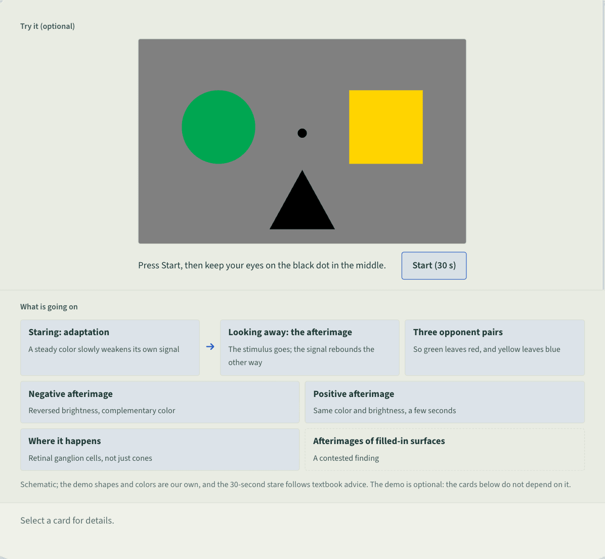
Press Start, then keep your eyes on the black dot in the middle.
What is going on
Schematic; the demo shapes and colors are our own, and the 30-second stare follows textbook advice. The demo is optional: the cards below do not depend on it.
Schematic. At the top, a gray box labeled try it (optional), with a black dot in the middle surrounded by a green circle, a yellow square and a black triangle, and a Start (30 s) button below. When the countdown ends the shapes disappear, leaving the gray field and the dot, with a note that the circle’s place may look reddish, the square’s bluish and the triangle’s lighter than its surroundings. Below, labeled what is going on, seven cards: staring, adaptation; looking away, the afterimage; three opponent pairs, red–green, yellow–blue, black–white; negative afterimage (reversed brightness, complementary color); positive afterimage (same color and brightness, a few seconds); where it happens (retinal ganglion cells, not just cones); afterimages of filled-in surfaces (a contested finding).
 Save as image (PNG)
This figure image is licensed CC BY 4.0: reuse it with credit and a link back to this page. How to reuse
Key points
- Opponent-process theory codes color in three pairs, red–green, yellow–blue and black–white: a cell excited by one member is inhibited by the other. Afterimage colors follow these pairings.[2]
- Positive afterimages also exist: rare, lasting a few seconds, with roughly the original color and brightness. Negative afterimages are more common, often more intense, and last longer.[1]
- Macaque recordings show retinal ganglion cells adapting slowly to a lasting color and rebounding when it stops; the authors trace everyday color afterimages to this, not to tired cones.[3]
- After fixating a neon color-spreading figure, observers also saw an afterimage of the perceptually filled-in surface, which the authors trace to cortical adaptation.[4] Others say retinal signals can explain it.[3]
An example
In a meeting, Lin stares at a bright green box on the projector screen for half a minute, then looks at the white wall. A faint red box of the same shape appears there and takes a moment to fade. After so much green, the wall looks tilted toward green’s opponent color, red.
Misreadings and limits
The demo is often used to show that perception “fills in” what is not there. What it shows directly is adaptation: after steady stimulation the visual system briefly swings the opposite way, so an afterimage’s color and brightness follow from the original.[1][3] Filling-in is a different effect; linking it to afterimages took specially designed figures.[4]
Sources
- 1 American Psychological Association (2018). Afterimage. APA Dictionary of Psychology (updated April 19, 2018). View source
- 2 Spielman, R. M., Jenkins, W. J., & Lovett, M. D. (2020). 5.3 Vision. Psychology 2e. OpenStax. View source
- 3 Zaidi, Q., Ennis, R., Cao, D., & Lee, B. (2012). Neural locus of color afterimages. Current Biology, 22(3), 220–224. doi:10.1016/j.cub.2011.12.021
- 4 Shimojo, S., Kamitani, Y., & Nishida, S. (2001). Afterimage of perceptually filled-in surface. Science, 293(5535), 1677–1680. doi:10.1126/science.1060161
Last checked against sources: October 28, 2026