Lobes of the brain
大脑叶
What are the lobes of the brain?
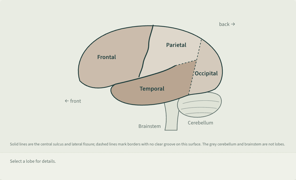
Textbooks usually divide the cortex of each cerebral hemisphere into four lobes: frontal, parietal, temporal and occipital.[1][2] The central sulcus separates frontal from parietal, the lateral (Sylvian) fissure sets the temporal lobe below both, and the parieto-occipital sulcus divides parietal from occipital.[2]
Solid lines are the central sulcus and lateral fissure; dashed lines mark borders with no clear groove on this surface. The grey cerebellum and brainstem are not lobes.
A side view of the left hemisphere, front to the left. The largest region, at the front, is the frontal lobe; behind and above it is the parietal lobe, separated by the central sulcus running down and forward from the top edge. The lateral fissure runs from low at the front up toward the back, with the temporal lobe below it. The occipital lobe is at the very back; its borders with the parietal and temporal lobes are dashed because no clear groove marks them on this surface. The cerebellum and brainstem are drawn in grey below and behind, marked as not lobes. Selecting a lobe shows its roles: frontal, movement, judgment, reasoning, impulse control and speech; parietal, touch, temperature, pain and combining the senses; temporal, hearing, memory, emotion and understanding language; occipital, vision.
 Save as image (PNG)
This figure image is licensed CC BY 4.0: reuse it with credit and a link back to this page. How to reuse
Key points
- Frontal lobe: the motor cortex plans and carries out voluntary movement, the prefrontal cortex supports judgment, reasoning and impulse control, and Broca’s area is essential for producing speech.[1][2]
- Parietal lobe: behind the central sulcus, the somatosensory cortex handles touch, temperature and pain from the opposite side of the body; right-sided damage can cause hemispatial neglect.[1][2]
- Temporal lobe: hearing, memory, emotion and some language. It holds the auditory cortex and Wernicke’s area for understanding speech; damage to the hippocampus inside it can cause anterograde amnesia.[1][2]
- Occipital lobe: at the very back, it holds the primary visual cortex, laid out to match positions in the visual field; damage to it on both sides can cause blindness.[1][2]
An example
At a crosswalk Lin hears someone call her name, turns to see a friend waving and waves back, while feeling her phone buzz in her pocket. Sound, sight, movement and touch are handled by areas in the temporal, occipital, frontal and parietal lobes, all working at once.[1][4]
Misreadings and limits
The claim that we use only 10% of our brains is false: imaging shows several areas at work in any activity.[4] “Left-brained” and “right-brained” personalities lack support too. Functions such as language do lean to one side, but a scan study of 1,011 people found no one with a stronger left- or right-brain network overall.[1][3]
Sources
- 1 Spielman, R. M., Jenkins, W. J., & Lovett, M. D. (2020). 3.4 The brain and spinal cord. Psychology 2e. OpenStax. View source
- 2 Javed, K., Reddy, V., & Lui, F. (2023). Neuroanatomy, cerebral cortex. StatPearls. StatPearls Publishing (last update July 25, 2023). View source
- 3 Nielsen, J. A., Zielinski, B. A., Ferguson, M. A., Lainhart, J. E., & Anderson, J. S. (2013). An evaluation of the left-brain vs. right-brain hypothesis with resting state functional connectivity magnetic resonance imaging. PLoS ONE, 8(8), e71275. doi:10.1371/journal.pone.0071275
- 4 BrainFacts/SfN (2010). Do you only use 10 percent of your brain?. BrainFacts.org, Society for Neuroscience. View source
Last checked against sources: October 27, 2026