Autonomic nervous system
自主神经系统
What is the autonomic nervous system?
The autonomic nervous system is the part of the peripheral nervous system that controls internal organs and glands, mostly outside voluntary control.[1] Its sympathetic division readies the body for stress and high arousal; its parasympathetic division, nicknamed “rest and digest,” returns it to routine.[1][2]
Heart rate (beats/min)
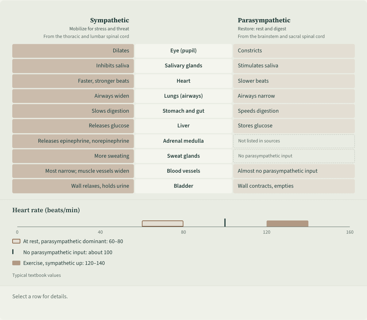
- At rest, parasympathetic dominant: 60–80
- No parasympathetic input: about 100
- Exercise, sympathetic up: 120–140
Typical textbook values
Three-column schematic. The middle column lists, top to bottom: eye, salivary glands, heart, lungs, stomach and gut, liver, adrenal medulla, sweat glands, blood vessels, bladder. The left column gives sympathetic effects: pupils dilate, saliva is inhibited, heart rate rises, airways widen, digestion slows, the liver breaks down glycogen and releases glucose, the adrenal medulla releases epinephrine and norepinephrine, sweating increases, most vessels narrow while those in skeletal muscle widen, the bladder wall relaxes. The right column gives parasympathetic effects: pupils constrict, saliva increases, heart rate falls, airways narrow, digestion speeds up, the liver stores glycogen; the adrenal row is blank, the sweat-gland and blood-vessel rows are marked as having no parasympathetic input; the bladder contracts. Below, a heart-rate scale marks three states: about 100 beats a minute without parasympathetic input, 60 to 80 at rest, 120 to 140 during exercise.
 Save as image (PNG)
This figure image is licensed CC BY 4.0: reuse it with credit and a link back to this page. How to reuse
Key points
- Sympathetic activity dilates the pupils, speeds the heart, widens the airways, slows digestion and makes the liver release glucose. Parasympathetic activity mostly does the reverse and also boosts salivation.[1][2]
- Most organs receive both. Left to itself the heart beats about 100 times a minute; the resting 60–80 reflects parasympathetic slowing, and sympathetic input speeds it up during exercise.[3]
- There are exceptions: sweat glands and most blood vessels are run by the sympathetic side alone, and the parasympathetic side has little effect on them.[2][3]
- In freezing animals both divisions are active at once, so heart rate may barely rise, hold steady or fall; in fight or flight, the parasympathetic brake on the heart is released.[4]
An example
As Ms. Wang steps up to present to her team, her palms sweat, her heart races, her mouth goes dry and her stomach knots. Back at her desk afterward, her heart slows and her stomach settles. Sympathetic activity dominates the first phase; in the second, the parasympathetic side takes over again.[1][2]
Misreadings and limits
A common picture treats the two divisions as a switch: sympathetic for stress and “bad,” parasympathetic for calm and “good.” In fact most organs receive both at once,[3] and the two can be active together, as in freezing.[4] Sympathetic arousal is a normal adaptation; constant, repeated stress is what is linked to health problems.[1]
Sources
- 1 Spielman, R. M., Jenkins, W. J., & Lovett, M. D. (2020). 3.3 Parts of the nervous system. Psychology 2e. OpenStax. View source
- 2 LeBouef, T., Yaker, Z., & Whited, L. (2023). Physiology, autonomic nervous system. StatPearls. StatPearls Publishing (last update May 1, 2023). View source
- 3 Betts, J. G., Young, K. A., Wise, J. A., Johnson, E., Poe, B., Kruse, D. H., Korol, O., Johnson, J. E., Womble, M., & DeSaix, P. (2022). 15.2 Autonomic reflexes and homeostasis. Anatomy and Physiology 2e. OpenStax. View source
- 4 Kozlowska, K., Walker, P., McLean, L., & Carrive, P. (2015). Fear and the defense cascade: Clinical implications and management. Harvard Review of Psychiatry, 23(4), 263–287. doi:10.1097/HRP.0000000000000065
Last checked against sources: October 27, 2026